Monday, March 27, 2023

Fumarate, mitochondria, and inflammation

Fumarate hydratase (FH), also known as fumarase, catalyses hydration of fumarate to malate and is found in mitochondria, where it performs in the Krebs cycle, and the cytosol, where it performs in the urea cycle and in catabolizing amino acids. It is encoded by homologous genes in bacteria, yeast, and eukaryotes. Fumarase deficiency in humans is involved in a range of symptoms from neurological abnormalities in newborns to tumors in adults.

These authors examined what happens immediately after loss of FH. They generated a mouse line with floxed Fh1 gene (homolog of the human FH) and crossed it with a line with ubiquitous, inducible, Cre recombinase (Rosa26). Induction of Cre caused the genetic loss of FH expression within 5 days and metabolic changes within 10 days (Fig 1). They found mitochondria swell and lose their genome (mtDNA) into the cytosol, where it triggers innate immunity (STING) and stimulates inflammation (RIG-I) including expression of interferon stimulated genes (ISGs). 

Treating cultured cells with a cell-permeable derivative of fumarate, monomethylfumarate (MMF), caused similar releases of mtDNA into the cytosol and expression of ISGs (Fig 3 Shown), strongly suggesting that the accumulation of fumarate causes the changes observed after loss of FH. They also show that mtDNA is released through ‘mitochondria-derived vesicles’ (MDVs), normally used to transport ‘content without affecting the integrity of the membranes’ (Fig 4), so the process occurs under some control. Finally, they show activation of DNA-sensor and innate immunity pathways in FH-deficient renal cancers, suggesting relevance for humans.

Fig 3a, Fumarate causes mitochondrial changes.  Overlay panels. Cells were treated for 8 days with solvent alone (vehicle) or with fumarate (200 or 400 uM MMF doses) then stained for DNA (green) and mitochondria (outer membrane protein TOM20, purple). Scale bars 10 um. Boxed area in left column expanded in right column.

One wonders what happens after the loss of other ancient and obscurely important cellular infrastructure.


Zecchini V, Paupe V, Herranz-Montoya I, Janssen J, Wortel IMN, Morris JL, Ferguson A, Chowdury SR, Segarra-Mondejar M, Costa ASH, Pereira GC, Tronci L, Young T, Nikitopoulou E, Yang M, Bihary D, Caicci F, Nagashima S, Speed A, Bokea K, Baig Z, Samarajiwa S, Tran M, Mitchell T, Johnson M, Prudent J, Frezza C. Fumarate induces vesicular release of mtDNA to drive innate immunity. Nature. 2023 Mar;615(7952):499-506. doi: 10.1038/s41586-023-05770-w. Epub 2023 Mar 8. PMID: 36890229; PMCID: PMC10017517.


Monday, February 27, 2023

Suppressing suppressors to improve lupus?

Lupus patients have more inflammation and more interferon type I (alphas, beta).  SLE monocytes have less NLRP12, which is part of certain inflammasome.and contributes to the activation of pro-inflammatory caspasesTLR7 is important in lupus.  The authors note that NLRP12 was ‘recently identified’ (citing a 2012 paper) as a negative regulator of TLR and NFkB activation. 

They found (Fig 1) NLRP12 expression is lower in lupus patients than in healthy controls and it is inversely correlated with type I interferon (IFN-a2) expression; (Fig 2) RUNX1 binding sequences in the NRLP12 promoter reduced expression of a reporter gene and CRISPR knockout of RUNX1 increased NLRP12 expression induced by IFN or virus; (Fig 3) there is more RUNX1 and less NLRP12 in monocytes from lupus patients (panels I, shown) and more RUNX1 binding to NLRP12 promoters in PBMC of lupus patients (panel J). (Fig 4) IFN-induced suppression of NLRP12 is mediated by histone acetylation; (Fig 5) RUNX1 reduces NLRP12, which increases IFN, which is (also) observed in SLE; (Fig 6) NLRP12-KO mice are more pro-inflammatory and (Fig 7 & 8) develop worse disease in mouse models of lupus (pristane injection or Fas-lpr).

Figure 3, panels I&J. Panel I immunoblot of proteins in lysates of CD14+ monocytes, quantified on right. Panel J ChIP (chromosome immunoprecipitation) of PBMC.

They do a very good job connecting pointillist dots.

Tsao YP, Tseng FY, Chao CW, Chen MH, Yeh YC, Abdulkareem BO, Chen SY, Chuang WT, Chang PC, Chen IC, Wang PH, Wu CS, Tsai CY, Chen ST. NLRP12 is an innate immune checkpoint for repressing IFN signatures and attenuating lupus nephritis progression. J Clin Invest. 2023 Feb 1;133(3):e157272. doi: 10.1172/JCI157272. PMID: 36719379.  

Monday, January 30, 2023

Can’t just “get over it”: Long COVID

Many people have experienced lingering problems after recovery from COVID infection, a condition known as long COVID or post-COVID.  A Norwegian study found that symptoms persisted for 6 months in most patients (189 of 312), including most young adults.  (‘Long COVID’ preferred because ‘post-COVID’ is ambiguous.) 

What is long COVID? There are many symptoms, led by fatigue, loss of smell/taste, breathlessness, and cognitive impairment, with no obvious common cause or relationship. Increased blood clotting (prothrombotic) has been suspected, suggested by the involvement of the receptor for the COVID-19 spike protein, ACE2, but strong evidence and mechanisms have been lacking.  

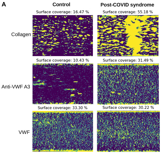
These authors tested the blood of 21 patients with post-COVID syndrome (PCS), averaging nearly 2 years after onset of infection. They modeled real blood flow through vessels by collecting blood samples (treated with the anti-coagulant citrate) and sending it through narrow tubes coated with particular proteins. They looked at the binding of platelets, which are abundant cellular products that initiate blood clots upon being triggered by binding collagen. They found striking increases in platelets binding to collagen (shown, Figure 1a top panel). Antibodies against von Willebrand factor (VWF) produced equivalent binding though apparently with different patterns (middle panels). 

Figure 1A: Binding of platelets (yellow) from healthy control blood (left) or PCS blood (right) to collagen (top), anti-VWF (middle), or VWF (bottom).
 

Although these intriguing findings await confirmation (by others) and follow-up, of course, they are of the utmost importance given the enormous impact of COVID.  

Constantinescu-Bercu A, Kessler A, de Groot R, Dragunaite B, Heightman M, Hillman T, Price LC, Brennan E, Sivera R, Vanhoorelbeke K, Singh D, Scully M. Analysis of thrombogenicity under flow reveals new insights into the prothrombotic state of patients with post-COVID syndrome. J Thromb Haemost. 2023 Jan;21(1):94-100. doi: 10.1016/j.jtha.2022.10.013. Epub 2022 Dec 22. PMID: 36695401; PMCID: PMC9773628.