Thursday, October 27, 2022

When viruses cohabit: Flu + RSV = Hybrid Frankenvirus

Experts worry that this winter might be made miserable by unwelcome visitors: something new, coronavirus variants, something flu (influenza A virus, IAV) and something blew, respiratory syncytial virus (RSV). What happens when somebody hosts IAV and RSV at the same time?

These investigators infected cultured human lung cells, A549 cells, and confirmed previous reports that coinfection reduces RSV but not IAV replication (Fig 1). Despite producing lower titer, they observed that infection with IAV appeared to increase the rate of coinfection by RSV.
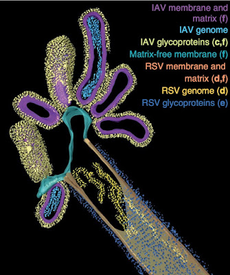
Fig 3b. Filament with features of IAV and RSV.

IAV and RSV are enveloped viruses that bud from the cell membrane with characteristic glycoproteins hemagglutinin (HA) and fusion (F), respectively. Having detected HA in areas of RSV budding from coinfected cells, the authors hypothesized that some virions would contain components of both viruses. Indeed, they observed many filaments, typical of RSV, with proteins from both viruses, albeit segregated (Fig 2a-e). A remarkable scanning electron micrograph appears to show hybrid viral particles (HVP) budding from the filaments (2f, red arrows). They analyzed the hybrid buds using cryo-ET and were able to ‘segment’ features of both viruses (shown, Fig 3b): mostly IAV virions budding from mostly RSV filaments. 

Amazing biology, but what does it mean clinically? The authors found that the hybrid virions contained IAV capable of infecting cells that had been depleted of their sialic acids, which bind HA, by treatment with neuraminidase (NA), Fig. 4-5). This could be an important mechanism widening the range of infected cells.

Haney J, Vijayakrishnan S, Streetley J, Dee K, Goldfarb DM, Clarke M, Mullin M, Carter SD, Bhella D, Murcia PR. Coinfection by influenza A virus and respiratory syncytial virus produces hybrid virus particlesNat Microbiol. 2022 Oct 24. doi: 10.1038/s41564-022-01242-5. Epub ahead of print. PMID: 36280786.

Friday, October 21, 2022

Bad news bears on life choices (vaccine hesitancy)

Vaccine hesitancy, a reluctance or refusal to be vaccinated, probably began when Jenner invented vaccination over two centuries ago. Vaccines have largely eliminated scourges such as smallpox and polio and greatly reduced the rates of other infectious diseases including influenza. Anti-vaccination (anti-vax) stances stem from small, well-established risks of side-effects (managed by a compensation program) and big, vague worries about unrelated, even disproven associations with other maladies. COVID-19 vaccines were developed rapidly and rushed into production, potentially raising valid safely concerns.  However, any valid concerns were allayed when the COVID-19 vaccines were tested and proven safe and effective (Walsh 2020). 

The cable television show Fox News Channel (FNC) amplified concerns about COVID-19 vaccines and downplayed their benefits.  This study of viewership and vaccination covered ~2,750 counties (out of ~3,000 total) in 47 (of 50) US states documents that FNC viewers refused COVID-19 vaccination more often than the viewers of its competitors Microsoft-National Broadcasting Company (MSNBC) or Cable News Network (CNN) (Figure 2, shown).

Figure 2. Effect of network viewership on weekly vaccination rates, 2021

A key question is whether FNC influenced its viewers to refuse vaccination (a cause) or rather were anti-vax viewers attracted to FNC’s messaging, a consequence of playing to its audience.  The investigators used positions in cable channel listing as ‘exogenous shifters’ of viewership (Martin & Yurukoglu 2017).  Viewers are induced into watching more or less of a channel by variation in its position up or down the listing (Fig S3).  They found that “exogenously higher FNC viewership due to channel position causes lower vaccine uptake”.  They show that hesitancy was raised by FNC but not by competitors MSNBC or CNN (Fig 1). Moreover, resistance to vaccination against COVID-19 but not seasonal flu… causal…. Using the channels’ position in the guides.

Their “results imply that watching one additional hour of [FNC] per week for the average household reduces the number of vaccinations by 0.35–0.76 per 100 people”, which would account for a lot of ‘excess deaths’ in many households. Not surprising when “vaccine bad” was said so much more often on FNC than the other channels (Fig S7)! Although they found that FNC’s influence was mostly on those under 65 years old, who are at lower risk severe disease, those younger people are reservoirs of virus for infecting older people. Data-driven lawyers representing survivors of FNC victims could bring class action lawsuits. 

Pinna, M., Picard, L. & Goessmann, C. Cable news andCOVID-19 vaccine uptake. Sci Rep 12, 16804 (2022). 

Tuesday, October 18, 2022

Lipid metabolism and dementia

About half the human brain mass is lipid. Several brain disorders are known to be caused by abnormal lipid metabolism, disruptions in the processes of making lipids and breaking them down. Second only to Alzheimer’s in prevalence is frontotemporal dementia (FTD), one form of which encompasses a range of social, behavioral, or language disorders (as opposed to memory or motor deficits seen in other disorders).  Several genes have been associated with FTD, foremost among them the conserved genes MAPT (tau), PSEN1 (presenilin), VCP (valosin containing protein) and GRN (granulin).


Granulins are a family of secreted, glycosylated peptides (A, B, C, etc.) cleaved from a single precursor (progranulin, PGRN), that are involved in a wide range of activities probably due to their roles in regulating protein lysosomal protein metabolism.  The authors of this study found that gangliosides (i.e., glycosphingolipids with attached sialic acids) are elevated in brains of granulin mutant mice (GRN R504X), which are analogous to the most prevalent granulin mutation in humans, R493X (substitution of the arginine normally at position 493 with a nonsense codon, resulting in a truncated protein). This mutation causes neuronal ceroid lipofuscinosis, a severe neurodevelopmental disease, in humans and neuroinflammation in mice (Jax).  The metabolic order in the ganglioside degradation pathway (Fig. 1a) is first disialylated GD1 (Fig. 1b, shown, rightmost plot, annotated with red #1) 👉 monosialylated GM1 (#2)  👉 GM2 (#3) 👉 GM3. Also, GD2 👉 GM3 (#3’) via an alternative pathway. Levels of precursor GD1 (#1) are significantly elevated in the brains of mice with heterozygous mutant granulin (Figure 1b: blue fill, Grn +/R493X) compared with normal granulin (Grn+/+, grey) but not in homozygous mutants (purple). This may suggest a feed-back mechanism that limits the accumulation of that metabolite. GM1, #2, is elevated in homozygous mutant brain compared to normal, with the heterozygous mutant intermediate. However, GM2, #3, is not significantly elevated in mutation-bearing mouse brains. The alternate pathway, #3’, shows elevated GD3, the precursor to GM3, in homozygous mutant brains.

They also analyzed the lipids in postmortem human brains of 12 GRN mutation-related FTD cases, 6 sporadic FTD cases, and 3 control normal subjects.  GD3 and GM1 are significantly elevated in GRN-related FTD cases (Fig 1c, blue columns). However, they both seem also elevated in non-GRN (sporadic) FTD cases (green). GD1 is significantly elevated in GRN-related but not -unrelated FTD.  

In a striking simplification, they tested the effects of removing the granulin precursor protein gene, PGRN (same as GRN), in HeLa cells (Fig 2). They found elevated GM2 in the deficient line (GRN-/-) that was reduced to normal levels by restoring granulin (GRN-/- + PGRN-addback).

What causes changes in the levels of gangliosides? Gangliosides are catabolized by lysosomal enzymes. However, those enzymes were not altered by GRN deficiency (Fig 3). An intermediate metabolite, bis(monoacylglycero)phosphate (BMP), which is crucial to ganglioside degradation, was found to be reduced by 50-60% in GRN-deficient HeLa cells and mouse brains, and ‘markedly’ in human brains of FTD cases, although again both GRN-related and sporadic cases (Fig 4d). The authors propose a model wherein “lysosomal granulin peptides maintain lysosomal function and homeostasis, including the levels of BMP, that are crucial for ganglioside catabolism”. Their results await confirmation by others and many details remain to be pursued further. One relatively simple aspect will be clarifying how the model accounts for autosomal dominance of GRN deficiency. Also worth noting is the proximity of GRN and the Alzheimer- and Parkinson-associated MAPT genes, within a million nucleotides in band 17q21.31. 

Boland S, Swarup S, Ambaw YA, Malia PC, Richards RC, Fischer AW, Singh S, Aggarwal G, Spina S, Nana AL, Grinberg LT, Seeley WW, Surma MA, Klose C, Paulo JA, Nguyen AD, Harper JW, Walther TC, Farese RV Jr. Deficiency of the frontotemporal dementia gene GRN results in gangliosidosis. Nat Commun. 2022 Oct 7;13(1):5924. doi: 10.1038/s41467-022-33500-9. PMID: 36207292; PMCID: PMC9546883.

Thursday, October 6, 2022

COVID restrictions – Moderation is good

In efforts to reduce the spread of COVID-19, billions of people around the world were subject to rules and laws governing their behavior. Among the various non-pharmaceutical interventions (NPIs), what worked? 


An influential early report analyzing data from 11 European countries during the first 4 months of the pandemic found that lockdowns reduced transmission rates (Rt values) significantly, ~80%, whereas other NPIs such as cancelling public events, school closure, encouraging social distancing, and self-isolation, resulted in less significant reductions (0-20%) (Fig 2, Flaxman).  The dramatic drop in infections after lockdown is so obvious that it required no modeling (Fig 1).  A similar study found the most effective NPIs for lowering cases were travel restrictions, school closures, and the partial lockdown (Cortis).  A related study of 19 NPIs during seasonal flu found that banning large gatherings was most effective in limiting transmission (Qiu).  These studies used epidemiological models that directly involve underlying mechanisms.


Data on cases, deaths, vaccinations, and tests, were obtained from the COVID-19 Data Hub (Guidotti).  NPI data were obtained from the Oxford Covid-19 Government Response Tracker (OxCGRT).  


In this report, the author analyzed data from 132 countries between Feb 2020 to April 2021, capturing 3 waves of infection, beginning March 202, July 2020, and January 2021.  An econometric model with 4 equations: C = cases growth rate, D= deaths growth rate, M = mobility, and p(SI) = probability of the assigned stringency intensity level was employed.  Stringency correlates inversely with nonresidential mobility (Fig 3).  He found that ‘unobserved variables’ influence the growth of cases and deaths (C and D) as well as the stringency (SI) of government policies. Medium-stringency measures greatly reduced case and death growth rates but, surprisingly, yet-more-stringent measures slightly increased them (Fig. 4, shown).   

Fig. 4 Case (panel a) and Death (panel b) growth rates vs NPI Stringency Index. 


Testing helped but contact tracing did little (Fig 5).  The benefits of reduced nonresidential mobility were outweighed by increased within-household transmission.  Various differences in culture, compliance, and enforcement of government imposed NPIs were acknowledged but not clearly managed. Even low levels of vaccination reduced Case and Death growth rates with nonlinear improvement anticipated. 


The findings disagree with previous reports that lockdowns are effective, concluding that "very stringent NPIs provide no further benefits over moderately stringent ones, and that less stringent NPIs function primarily as signals for significant voluntary changes in citizens’ behavior.". Such analyses are crucial for designing effective, results-driven policies and for persuading people to comply.  


Spiliopoulos L. On the effectiveness of COVID-19 restrictions and lockdowns: Pan metron ariston. BMC Public Health. 2022 Oct 1;22(1):1842. doi: 10.1186/s12889-022-14177-7. PMID: 36183075; PMCID: PMC9526209.