Wednesday, September 28, 2022

Antibody specificities are Gut reactions

Germ-free (GF) mice have undeveloped immune systems and practically no antibodies.  How do microbes in the gut (gastro-intestinal tract) stimulate immunity?  This group looked at the specificity of antibodies that develop after GF mice are colonized by individual bacterial species and strains (monocolonization).  They used 8 strains of bacteria to inoculate GF mice and, after 3 weeks, analyzed the specificity of antibodies produced in the gut (recovered from fecal matter) and blood, focusing on the IgA isotype that protects mucosal surfaces.  

Not surprisingly, the antibodies tend to bind specifically to those strains of microbes against which they were stimulated (shown, Figure 1, panel A). They also found that mice monocolonized from birth produce more IgA reactive with that species (termed ‘self’) than newly-introduced species. They make a point about IgA being able to ‘aggregate pathogenic bacteria’ and ‘selectively coat disease-associated bacteria’ but it is unclear how IgA itself could distinguish dangerous from benign and anyway they tested only benign bacteria.  

A small panel (29) of monoclonal IgA antibodies cloned from gut tissues of monocolonized mice also showed species specificity. Finally, they showed that monoclonal IgA antibodies with specific binding activity could be detected in the feces of mice that had been force-fed the IgA 3 hours previously, suggesting a targeted, potential therapy (e.g., against the human pathogen Clostridioides difficile). 

Yang C, Chen-Liaw A, Spindler MP, Tortorella D, Moran TM, Cerutti A, Faith JJ. Immunoglobulin A antibody composition is sculpted to bind the self gut microbiome. ScienceImmunology. 2022 Jul 15;7(73) 

Wednesday, September 21, 2022

Covid Vax vs Variants

The first RNA vaccines against Covid were based on the Surface, S or “Spike” protein from the reference genome of SARS-CoV-2, Wuhan-Hu-1, published in January 2020.  Since then, more-infectious variants have emerged; one, D614G, dominated but did not escape vaccine protection probably because its characteristic mutation is located outside the Receptor Binding Domain (RBD, 319-541). In the past year, delta variants were overtaken by omicron variants, which have numerous changes in the RBD, raising concern that they escape current RNA vaccines. 

Neutralization assays measure the ability of blood-borne antibodies to block viral infection of cells grown in a culture dish; they are thought to provide valid measurements of protection. These authors previously tested Covid neutralization with blood sera from 15 health care workers, 4 vaccinated with Moderna (mRNA-1273) and 11 with Pfizer-BioNTech (BNT162b2). They confirmed that a third dose (second booster) increased the neutralization activity (titer) against all strains, albeit 3-4 times weaker against subvariants compared with the ancestral version. Moreover, they showed the neutralization titers induced by 3 doses of the vaccines, and therefore presumably levels of protection provided, approximated those found in convalescent (sick) Covid patients (Fig 1 panel B vs C and D). 

In this letter, they studied 46 health care workers, 24 vaccinated and boosted with Moderna and 22 with Pfizer-BioNTech. Fourteen of the cohort were infected during the follow-up year. Figure S3 in the supplementary data confirm the protection provided by a third dose (second booster). They found that neutralization titers against all strains declined over time.  Titers from vaccinated people remained within a substantial fraction of those from infected people (shown, dashed vs solid lines). 

How much protection -- neutralization titer -- is enough? A paper published last year analyzing 7 different vaccines reported that neutralization titers are predictive of protection.  Neutralization titers of 20% of convalescent levels protected on average half of people against detectable infection; levels at 3% convalescent protected against severe Covid. Taken together, these results seem to support the value of the ‘old’ vaccines against the newer variants. 

Qu P, Faraone JN, Evans JP, Zheng YM, Yu L, Ma Q, Carlin C, Lozanski G, Saif LJ, Oltz EM, Gumina RJ, Liu SL. Durability of Booster mRNA Vaccine against SARS-CoV-2 BA.2.12.1, BA.4, and BA.5 Subvariants. N Engl J Med. 2022 Sep 7. 

Tuesday, September 13, 2022

Obesity and severe COVID

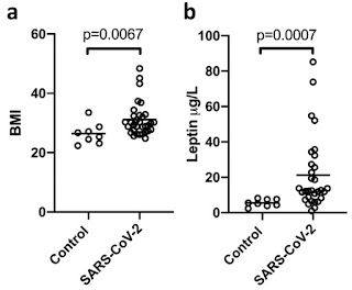
Early in the COVID-19 pandemic, it was recognized that obesity seemed to be related to respiratory failure, or severe acute respiratory syndrome (SARS). These researchers tested whether leptin, a cytokine produced by fat cells in the gut and working on brain cells to influence hunger, might be correlated with risk, perhaps a biomarker of risk. 

They compared 31 obese COVID patients on ventilators with 8 non-infected, non-obese critically ill patients.  They found much higher levels of leptin in the patients with COVID (averages 21 vs 6 ug/l, with very good statistical significance, p = .0007).  The individual measurements overlap (shown), so leptin is not ‘the’ biomarker but clearly related. Whether related as a cause or consequence only a prospective trial could test rigorously. 

Figure 2 detail: BMI of patients panel A and Leptin levels panel B.  In each panel, ‘control’ critically ill patients, left, COVID patients right. 


The authors hypothesized that elevated leptin causes a ‘hyper immune’ state, especially stimulating lung epithelial cells.  They noted similar observation previously published for influenza and MERS. Since this paper was published, several groups have reported similar findings 2020 paper that was largely replicated (see review, which cites 3 later papers).  Ironically, shutdowns that have been effective in protecting many people from infection have also increased sedentary lifestyles, BMI, and risk. 


van der Voort PHJ, Moser J, Zandstra DF, Muller Kobold AC, Knoester M, Calkhoven CF, Hamming I, van Meurs M. Leptin levels in SARS-CoV-2 infection related respiratory failure: A cross-sectional study and a pathophysiological framework on the role of fat tissue. Heliyon. 2020 Aug;6(8):

Wednesday, September 7, 2022

A kidney ‘punch’ can tell much

renal biopsy is when a small piece of a patient's kidney is removed for analysis, usually by inserting a needle through the skin and into the kidney. Though invasive, the clinical value can be high for helping decide how to treat lupus or transplanted patients.  How to maximize that value? 

In this study, Clark and colleagues applied computer imaging techniques to answer why only about half of lupus patients with inflamed kidneys (nephritis) proceed to kidney failure (end stage renal disease, ESRD) and lose their kidney. They supposed there were subtle differences in the “frequency and organization of principal cellular effectors” between those patients who did, or didn’t, progress. They obtained biopsies from a cohort of 55 well-characterized lupus patients, of which 19 progressed to ESRD. They labeled very thin slices (sections) of kidney with 6 markers (CD3, CD4, CD20, CD11c, BDCA2, and DAPI) and counted several types of immune cells using confocal microscopy and deep learning analysis. 

Figure 2: left panel (H) are CD20+ B cells, right panel (I are CD3+CD4- T cells, (probably CD8+?).


They found a remarkably clear distinction: those with few B cells and many CD4- T cells, probably CD8+, often progress to ESRD (shown in red, panels H and I from figure 2).  Patients with many B cells and few CD4- T cells do not progress to ESRD (blue).  In the discussion, the authors mention that some clinical trials have targeted exactly those cells that this study indicate may be protective (B cells) or innocuous (CD4+ T cells).  A validated "identification, friend or foe" (IFF) system seems a good principle before aiming and firing.  


Abraham R, Durkee MS, Ai J, Veselits M, Casella G, Asano Y, Chang A, Ko K, Oshinsky C, Peninger E, Giger ML, Clark MR. Specific in situ inflammatory states associate with progression to renal failure in lupus nephritis. J Clin Invest. 2022 Jul 1;132(13)