Friday, February 28, 2025

Autoantibodies cause some Long-Covid symptoms

Experience with other viral infections suggests that increased autoimmunity following infection with COVID-19 might contribute to the symptoms of Post-Acute Sequelae of SARS-CoV-2 infection (PASC), more commonly known as Long COVID (LCOVID).  LCOVID is characterized by the presence of autoantibodies of diverse specificities and clinical improvement is correlated with their decline.  Immunoglobulin G (IgG) antibodies from fibromyalgia patients has been shown to cause pain in mice.  Therefore, this group tested the effects on mice of IgG antibodies from LCOVID patients. 

Most LCOVID patients’ plasma showed reduced IFN-gamma (contrast this report) and many showed elevated Glial Fibrillary Acidic Protein (GFAP, which is neuroprotective); other measured proteins were unchanged (Fig 1).  Patient sera were separated into 3 groups based on neuronal damage and astroglial activation markers (Long Covid-1, LC-1), and type I IFNs (LC-2 higher IFNa2a, beta, LC-2 lower).  Principal component analysis of sera proteome signatures confirmed groupings (Figs 3 & 4).  

 IgG from patients with elevated neuronal proteins (LC-1) or leukocyte activation markers (LC-3) caused sensory hypersensitivity (Fig 5, shown; LC1 red, LC3 yellow ).  They claim that IgG from patients with higher muscle proteins also reduced locomotor movements in mice, though the extent appears small (Fig 6).  This careful study is an important guide to such analyses.  
Hung-Jen Chen, Brent Appelman, Hanneke Willemen, Amelie Bos, Judith Prado, Chiara. E. Geyer, Patrícia Silva Santos Ribeiro, Sabine Versteeg, Mads Larsen, Eline Schüchner, Marije K. Bomers, Ayesha H.A. Lavell, Amsterdam UMC COVID-19 biobank, Braeden Charlton, Rob Wüst, W. Joost Wiersinga, Michèle van Vugt1, Gestur Vidarsson, Niels Eijkelkamp, Jeroen den Dunnen . Transfer of IgG from Long COVID patients induces symptomology in mice. BioRxiv, May 31. 2024.


Monday, March 27, 2023

Fumarate, mitochondria, and inflammation

Fumarate hydratase (FH), also known as fumarase, catalyses hydration of fumarate to malate and is found in mitochondria, where it performs in the Krebs cycle, and the cytosol, where it performs in the urea cycle and in catabolizing amino acids. It is encoded by homologous genes in bacteria, yeast, and eukaryotes. Fumarase deficiency in humans is involved in a range of symptoms from neurological abnormalities in newborns to tumors in adults.

These authors examined what happens immediately after loss of FH. They generated a mouse line with floxed Fh1 gene (homolog of the human FH) and crossed it with a line with ubiquitous, inducible, Cre recombinase (Rosa26). Induction of Cre caused the genetic loss of FH expression within 5 days and metabolic changes within 10 days (Fig 1). They found mitochondria swell and lose their genome (mtDNA) into the cytosol, where it triggers innate immunity (STING) and stimulates inflammation (RIG-I) including expression of interferon stimulated genes (ISGs). 

Treating cultured cells with a cell-permeable derivative of fumarate, monomethylfumarate (MMF), caused similar releases of mtDNA into the cytosol and expression of ISGs (Fig 3 Shown), strongly suggesting that the accumulation of fumarate causes the changes observed after loss of FH. They also show that mtDNA is released through ‘mitochondria-derived vesicles’ (MDVs), normally used to transport ‘content without affecting the integrity of the membranes’ (Fig 4), so the process occurs under some control. Finally, they show activation of DNA-sensor and innate immunity pathways in FH-deficient renal cancers, suggesting relevance for humans.

Fig 3a, Fumarate causes mitochondrial changes.  Overlay panels. Cells were treated for 8 days with solvent alone (vehicle) or with fumarate (200 or 400 uM MMF doses) then stained for DNA (green) and mitochondria (outer membrane protein TOM20, purple). Scale bars 10 um. Boxed area in left column expanded in right column.

One wonders what happens after the loss of other ancient and obscurely important cellular infrastructure.


Zecchini V, Paupe V, Herranz-Montoya I, Janssen J, Wortel IMN, Morris JL, Ferguson A, Chowdury SR, Segarra-Mondejar M, Costa ASH, Pereira GC, Tronci L, Young T, Nikitopoulou E, Yang M, Bihary D, Caicci F, Nagashima S, Speed A, Bokea K, Baig Z, Samarajiwa S, Tran M, Mitchell T, Johnson M, Prudent J, Frezza C. Fumarate induces vesicular release of mtDNA to drive innate immunity. Nature. 2023 Mar;615(7952):499-506. doi: 10.1038/s41586-023-05770-w. Epub 2023 Mar 8. PMID: 36890229; PMCID: PMC10017517.


Monday, February 27, 2023

Suppressing suppressors to improve lupus?

Lupus patients have more inflammation and more interferon type I (alphas, beta).  SLE monocytes have less NLRP12, which is part of certain inflammasome.and contributes to the activation of pro-inflammatory caspasesTLR7 is important in lupus.  The authors note that NLRP12 was ‘recently identified’ (citing a 2012 paper) as a negative regulator of TLR and NFkB activation. 

They found (Fig 1) NLRP12 expression is lower in lupus patients than in healthy controls and it is inversely correlated with type I interferon (IFN-a2) expression; (Fig 2) RUNX1 binding sequences in the NRLP12 promoter reduced expression of a reporter gene and CRISPR knockout of RUNX1 increased NLRP12 expression induced by IFN or virus; (Fig 3) there is more RUNX1 and less NLRP12 in monocytes from lupus patients (panels I, shown) and more RUNX1 binding to NLRP12 promoters in PBMC of lupus patients (panel J). (Fig 4) IFN-induced suppression of NLRP12 is mediated by histone acetylation; (Fig 5) RUNX1 reduces NLRP12, which increases IFN, which is (also) observed in SLE; (Fig 6) NLRP12-KO mice are more pro-inflammatory and (Fig 7 & 8) develop worse disease in mouse models of lupus (pristane injection or Fas-lpr).

Figure 3, panels I&J. Panel I immunoblot of proteins in lysates of CD14+ monocytes, quantified on right. Panel J ChIP (chromosome immunoprecipitation) of PBMC.

They do a very good job connecting pointillist dots.

Tsao YP, Tseng FY, Chao CW, Chen MH, Yeh YC, Abdulkareem BO, Chen SY, Chuang WT, Chang PC, Chen IC, Wang PH, Wu CS, Tsai CY, Chen ST. NLRP12 is an innate immune checkpoint for repressing IFN signatures and attenuating lupus nephritis progression. J Clin Invest. 2023 Feb 1;133(3):e157272. doi: 10.1172/JCI157272. PMID: 36719379.  

Monday, January 30, 2023

Can’t just “get over it”: Long COVID

Many people have experienced lingering problems after recovery from COVID infection, a condition known as long COVID or post-COVID.  A Norwegian study found that symptoms persisted for 6 months in most patients (189 of 312), including most young adults.  (‘Long COVID’ preferred because ‘post-COVID’ is ambiguous.) 

What is long COVID? There are many symptoms, led by fatigue, loss of smell/taste, breathlessness, and cognitive impairment, with no obvious common cause or relationship. Increased blood clotting (prothrombotic) has been suspected, suggested by the involvement of the receptor for the COVID-19 spike protein, ACE2, but strong evidence and mechanisms have been lacking.  

These authors tested the blood of 21 patients with post-COVID syndrome (PCS), averaging nearly 2 years after onset of infection. They modeled real blood flow through vessels by collecting blood samples (treated with the anti-coagulant citrate) and sending it through narrow tubes coated with particular proteins. They looked at the binding of platelets, which are abundant cellular products that initiate blood clots upon being triggered by binding collagen. They found striking increases in platelets binding to collagen (shown, Figure 1a top panel). Antibodies against von Willebrand factor (VWF) produced equivalent binding though apparently with different patterns (middle panels). 

Figure 1A: Binding of platelets (yellow) from healthy control blood (left) or PCS blood (right) to collagen (top), anti-VWF (middle), or VWF (bottom).
 

Although these intriguing findings await confirmation (by others) and follow-up, of course, they are of the utmost importance given the enormous impact of COVID.  

Constantinescu-Bercu A, Kessler A, de Groot R, Dragunaite B, Heightman M, Hillman T, Price LC, Brennan E, Sivera R, Vanhoorelbeke K, Singh D, Scully M. Analysis of thrombogenicity under flow reveals new insights into the prothrombotic state of patients with post-COVID syndrome. J Thromb Haemost. 2023 Jan;21(1):94-100. doi: 10.1016/j.jtha.2022.10.013. Epub 2022 Dec 22. PMID: 36695401; PMCID: PMC9773628.  


Monday, December 26, 2022

Identifying Mitochondrial Functions by ‘Multiomic’ Profiling

Mitochondria make ATP through oxidative phosphorylation, thus providing energy for nearly all cellular functions. Many human disorders are attributed to mitochondrial dysfunction. Their functions seem narrow and their genomes, known for decades, were reduced to encoding a mere 13 proteins after transferring most to the nuclear genome. However, these authors note that ‘hundreds of mitochondrial proteins lack clear functions’. They previously (Stefely 2016) applied mass spectroscopy (MS) ‘multiomics’ to assign functions to mitochondrial uncharacterized (x) proteins (MXPs) in yeast. Here, they generated using CRISPR over 200 knockout (KO) cell lines, targeting 50 nuclear genes encoded MXPs plus 66 with known functions, and assessed in each line over 8,000 proteins, over 3,000 lipids and over 200 metabolites by MS (epic undertaking!). They found high reproducibility and dynamic range, with ‘many molecules showing regulation over 3-4 orders of magnitude’.

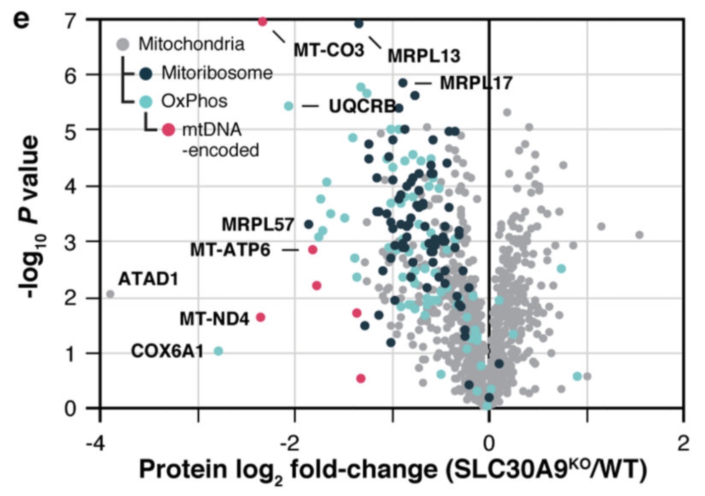
Fig 2e. Relative protein abundance in SLC30A9 KO cells compared to WT (“wild type”, i.e., normal) cells versus statistical significance with noted mitochondrial ribosome (black), OxPhos (blue), and mtDNA-encoded (red) proteins.

Some assessments confirmed expectations or were mild surprises, e.g., the importance of ALDH18A1 or NADK1 in proline synthesis (Fig 2a). Others revealed ‘new biology’, such as a key role for the putative zinc transporter SLC30A9 in mitochondrial ribosome and OxPhos proteins (Fig 2e, shown). They also found that one ’upstream (open) reading frame’ (PYURF) is a chaperone essential for complex I and coQ synthesis, linked a transporter (SLC30A9) to ribosomes, and found a second gene (RAB5IF) contributing to developmental disorders. They offer their “8.3 million distinct biomolecule measurements” online to help others ascribe additional functions, a promising resource.

Rensvold JW, Shishkova E, Sverchkov Y, Miller IJ, Cetinkaya A, Pyle A, Manicki M, Brademan DR, Alanay Y, Raiman J, Jochem A, Hutchins PD, Peters SR, Linke V, Overmyer KA, Salome AZ, Hebert AS, Vincent CE, Kwiecien NW, Rush MJP, Westphall MS, Craven M, Akarsu NA, Taylor RW, Coon JJ, Pagliarini DJ. Defining mitochondrial protein functions through deep multiomic profiling. Nature. 2022 Jun;606(7913):382-388. doi: 10.1038/s41586-022-04765-3. Epub 2022 May 25. PMID: 35614220; PMCID: PMC9310563.
NB PubPeer comment raises concerns regarding the methods, interpretations, and conclusions. 

Friday, December 16, 2022

Seasonal flu vaccination: short term protection, long term risk?

Influenza (flu) sickens millions and kills many thousands of Americans each year. Vaccination is intended to reduce the number cases and severity of illness. The flu virus changes its coat proteins each year, a ‘shape-shifting’ behavior that challenges the timely production of vaccines that are effective against flu variants. The US CDC evaluates vaccine effectiveness (VE) in thousands of outpatient respiratory illness patients, usually finding substantial vaccine protection with little risk Consequently, its Advisory Committee on Immunization Practices (ACIP) as well as the World Health Organization (WHO) recommend annual immunization of everyone over 6 months old unless contraindicated.
 
Here, the authors surveyed vaccination over 10 years among several thousand Japanese school children and several hundred school staff adults. Of the many forms of flu vaccine, Japan uses a quadrivalent (4 strains; trivalent prior to 2014) “split” vaccine based on influenza hemagglutinin (HA). “Split” vaccine means the virus was ‘disrupted’ by detergent (the equivalent of ‘heat killed’; viruses aren’t alive). The flu viruses used to prepare the vaccine are grown in eggs; alternative quadrivalent preparations are available for those with allergies to eggs, including Flublok and Flucelvax.

They found that morbidity was reduced in vaccinated elementary school students but elevated in middle school students (Fig 1). Most people who had been vaccinated ‘from infancy’ were also vaccinated in the 2019-2020 season (Fig. 2). Moreover, they “found that morbidity was significantly higher among elementary (P < 0.001) and middle (P < 0.05) school students who had been vaccinated since infancy than among those who had not been vaccinated since infancy” (Figure 3, shown).

Fig. 3 Relationship between morbidity and vaccination from infancy.

These data are self-reported via questionnaires, and therefore extra subjective. The authors propose no mechanism for how annual vaccination could cause increased morbidity. It seems probable that the association is not direct, not causative, but indirect through other behavioral or health status factors. This is an intriguing finding that should be analyzed and study that should be repeated.

Kajiume T, Mukai S, Toyota N, Kanazawa I, Kato A, Akimoto E, Shirakawa T. Effectiveness of seasonal influenza vaccine in elementary and middle schools: a 10-year follow-up investigation. BMC Infect Dis. 2022 Dec 6;22(1):909. doi: 10.1186/s12879-022-07898-y. PMID: 36474168; PMCID: PMC9724312.

Wednesday, November 16, 2022

Protein folding by AI: wrinkles

Tech giants Alpha and Meta (Google and Facebook) applied their Artificial Intelligence (AI) to fold proteins computationally, predicting 3-dimensional shapes from the 1-dimensional sequence data. Meta’s paper is still paywalled (preprint) but AlphaFold’s Nature papers from last year are available (Jumper, Tunyasuvunakool). 

The AlphaFold authors noted that the ~100,000 protein structures determined by conventional experimental means are a small portion of the “billions” extant in nature. Previous approaches “focus on either the physical interactions or the evolutionary history”, which they say relies on the availability of close homologues or works for (only) a few, small proteins and is otherwise “computationally intractable” (too hard). They evaluated in the 87 protein domains comprising the 14th Critical Assessment of (protein) Structure Prediction (CASP14) dataset, structures not yet deposited in the public Protein Data Bank (PDB). This permits a ‘blind’ (apriori) comparison of AI methods, by comparing their predictions with the newly-solved structures. 

Fig 1. a. Scores. b. Backbone. c. Side chains
 By this measure, AlphaFold is much better than its competitors (Fig 1a, shown, predicted vs experimental). It gains accuracy on backbone and side chains (1b, c) “by incorporating novel neural network architectures and training procedures based on the evolutionary, physical and geometric constraints of protein structures”. Using “multiple sequence alignments (MSAs) and pairwise” comparisons, it “predicts the 3D coordinates of all heavy atoms for a given protein using the primary amino acid sequence and aligned sequences of homologues as inputs”. So give it a bunch of similar sequences and structures and voila! it gives the ‘new’ one. Thy describe the process and you can download to code to inspect, modify, run yourself (open source).  

While impressive, this is a very constrained set of structures, nothing justifying the claims made in the popular press of solving all proteins. To be comprehensive, it seems that AI will have to consider biology, implement means of including the amino-terminal-first synthesis, nucleation, domain folding, insertion into a membrane, and above all interaction with chaperone proteins. 

Jumper J, Evans R, Pritzel A, Green T, Figurnov M, Ronneberger O, Tunyasuvunakool K, Bates R, Žídek A, Potapenko A, Bridgland A, Meyer C, Kohl SAA, Ballard AJ, Cowie A, Romera-Paredes B, Nikolov S, Jain R, Adler J, Back T, Petersen S, Reiman D, Clancy E, Zielinski M, Steinegger M, Pacholska M, Berghammer T, Bodenstein S, Silver D, Vinyals O, Senior AW, Kavukcuoglu K, Kohli P, Hassabis D. Highly accurate protein structure prediction with AlphaFold. Nature. 2021 Aug;596(7873):583-589. doi: 10.1038/s41586-021-03819-2. Epub 2021 Jul 15. PMID: 34265844; PMCID: PMC8371605.

Saturday, November 12, 2022

Leptin boosts protective vaccine responses

Follicular helper T cells (Tfh) support B cell development and production of antibodies, essential for a protective vaccination response. Metabolism has been linked to T cell development and the metabolic hormone leptin varies up to 10-fold among healthy people. Here, the investigators asked whether leptin levels might influence T cell development and contribute to variability in vaccine responses. 

Within a cohort of 76 healthy adults, they found non-responders to influenza vaccination had on average 2.5-fold lower serum leptin levels, with non-responders 10-fold more frequent in the low leptin group (fig 1ab). Tfh counts correlate with leptin levels (fig 2). Similar observations were made among older flu vaccine recipients (age >64 yr) and young Hepatitis B vaccine (HBV) recipients. Adding leptin to T cells cultured in vitro increased Tfh markers and production of IL-21 (fig 2e). 

In mice, they found leptin in areas of B cell development and leptin receptors on Tfh cells. Leptin receptor deficiency reduced antibody responses (fig 3b, c) and (consequently) allowed viral growth (panel a) in mice infected with H1N1 influenza. Tfh in leptin-receptor-deficient mice produced less IL-21 (fig 5b) and supplemental IL-21 restored most antibody production (fig 5a). IL-21 production is abrogated in T cells lacking STAT3 (fig 5g), strongly supporting a mechanism involving STAT3 and IL-21. 

Fig 7. Leptin protects from fasting-induced susceptibility to influenza. 


They could transiently reduce serum leptin levels by ‘fasting’ (starving) mice on alternate days 5 to 15 days after infection with influenza (Fig 7a, shown above). This timing chosen to avoid interfering with T cell priming (d 0-5) and focus on peak Tfh development (starting d5). Supplemental leptin protected against influenza (panel b), underscoring the significance of this pathway.

Deng J, Chen Q, Chen Z, Liang K, Gao X, Wang X, Makota FV, Ong HS, Wan Y, Luo K, Gong D, Yu X, Camuglia S, Zeng Q, Zhou T, Xue F, He J, Wei Y, Xiao F, Ma J, Hill DL, Pierson W, Nguyen THO, Zhou H, Wang Y, Shen W, Sun L, Li Z, Xia Q, Qian K, Ye L, Rockman S, Linterman MA, Kedzierska K, Shen N, Lu L, Yu D. The metabolic hormone leptin promotes the function of TFH cells and supports vaccine responses. Nat Commun. 2021 May 24;12(1):3073. doi: 10.1038/s41467-021-23220-x. PMID: 34031386; PMCID: PMC8144586.

Wednesday, November 2, 2022

Physical activity increases gut bacteria diversity

Previous work established associations in humans between physical activity and reduced obesity, reduced mortality, and improved cardiovascular health. Physical activity has been also associated with the microbiome in animals. Here, the relationship between physical activity and microbiome in humans was investigated.

The authors studied a cohort of 720 adults, citizens of Wisconsin, average age 55 years, 83% White, 10% Black, 42% male. Gut microbial, (bacterial) composition was assessed using sequencing the V3-V4 region of 16S rRNA extracted from stool samples. Note this is only a subset of the ‘microbiome’, not include non-bacterial components such as fungi, viruses, etc. 

They monitored physical activity using accelerometers worn on the hip (activity) or wrist (sleep). Participants also self-reported whether in a typical week they walked or biked at least 10 minutes continuously to get around. Those who responded ‘yes’ were classified as participating in ‘active transportation’. Note this is a threshold of less than half a mile a week, walking only about 100 m per day. 

Table 2. Linear mixed effects models (adjusted for characteristics, Table 1). CI, confidence interval; SD, standard deviation; MVPA, moderate to vigorous physical activity; ** p<0.05. *** p < 0.01.
They identified 865 unique bacterial taxa, largely encompassed by about 20 abundant phyla (Fig 1). They observed no change in bacterial diversity in participants who engaged in moderate-to-vigorous activity (line 2, Table 2, shown) or active transportation (line 3). However, when they analyzed those participants who engaged in higher levels of active transportation, at least 1 standard deviation (SD) above the average, they observed significant increases in bacterial diversity (line 4).

They also found the abundance of an unknown family from order Clostridiales was associated with increased weekly MVPA minutes. They conclude that their results “point to a potential pathway by which the gut micro- biota may be linked to physical activity and other well established health benefits”.

Holzhausen EA, Malecki KC, Sethi AK, Gangnon R, Cadmus-Bertram L, Deblois CL, Suen G, Safdar N, Peppard PE. Assessing the relationship between physical activity and the gut microbiome in a large, population-based sample of Wisconsin adults. PLoS One. 2022 Oct 26;17(10):e0276684. doi: 10.1371/journal.pone.0276684. PMID: 36288361; PMCID: PMC9605031.

Thursday, October 27, 2022

When viruses cohabit: Flu + RSV = Hybrid Frankenvirus

Experts worry that this winter might be made miserable by unwelcome visitors: something new, coronavirus variants, something flu (influenza A virus, IAV) and something blew, respiratory syncytial virus (RSV). What happens when somebody hosts IAV and RSV at the same time?

These investigators infected cultured human lung cells, A549 cells, and confirmed previous reports that coinfection reduces RSV but not IAV replication (Fig 1). Despite producing lower titer, they observed that infection with IAV appeared to increase the rate of coinfection by RSV.

Fig 3b. Filament with features of IAV and RSV.

IAV and RSV are enveloped viruses that bud from the cell membrane with characteristic glycoproteins hemagglutinin (HA) and fusion (F), respectively. Having detected HA in areas of RSV budding from coinfected cells, the authors hypothesized that some virions would contain components of both viruses. Indeed, they observed many filaments, typical of RSV, with proteins from both viruses, albeit segregated (Fig 2a-e). A remarkable scanning electron micrograph appears to show hybrid viral particles (HVP) budding from the filaments (2f, red arrows). They analyzed the hybrid buds using cryo-ET and were able to ‘segment’ features of both viruses (shown, Fig 3b): mostly IAV virions budding from mostly RSV filaments. 

Amazing biology, but what does it mean clinically? The authors found that the hybrid virions contained IAV capable of infecting cells that had been depleted of their sialic acids, which bind HA, by treatment with neuraminidase (NA), Fig. 4-5). This could be an important mechanism widening the range of infected cells.

Haney J, Vijayakrishnan S, Streetley J, Dee K, Goldfarb DM, Clarke M, Mullin M, Carter SD, Bhella D, Murcia PR. Coinfection by influenza A virus and respiratory syncytial virus produces hybrid virus particlesNat Microbiol. 2022 Oct 24. doi: 10.1038/s41564-022-01242-5. Epub ahead of print. PMID: 36280786.

Friday, October 21, 2022

Bad news bears on life choices (vaccine hesitancy)

Vaccine hesitancy, a reluctance or refusal to be vaccinated, probably began when Jenner invented vaccination over two centuries ago. Vaccines have largely eliminated scourges such as smallpox and polio and greatly reduced the rates of other infectious diseases including influenza. Anti-vaccination (anti-vax) stances stem from small, well-established risks of side-effects (managed by a compensation program) and big, vague worries about unrelated, even disproven associations with other maladies. COVID-19 vaccines were developed rapidly and rushed into production, potentially raising valid safely concerns.  However, any valid concerns were allayed when the COVID-19 vaccines were tested and proven safe and effective (Walsh 2020). 

The cable television show Fox News Channel (FNC) amplified concerns about COVID-19 vaccines and downplayed their benefits.  This study of viewership and vaccination covered ~2,750 counties (out of ~3,000 total) in 47 (of 50) US states documents that FNC viewers refused COVID-19 vaccination more often than the viewers of its competitors Microsoft-National Broadcasting Company (MSNBC) or Cable News Network (CNN) (Figure 2, shown).

Figure 2. Effect of network viewership on weekly vaccination rates, 2021

A key question is whether FNC influenced its viewers to refuse vaccination (a cause) or rather were anti-vax viewers attracted to FNC’s messaging, a consequence of playing to its audience.  The investigators used positions in cable channel listing as ‘exogenous shifters’ of viewership (Martin & Yurukoglu 2017).  Viewers are induced into watching more or less of a channel by variation in its position up or down the listing (Fig S3).  They found that “exogenously higher FNC viewership due to channel position causes lower vaccine uptake”.  They show that hesitancy was raised by FNC but not by competitors MSNBC or CNN (Fig 1). Moreover, resistance to vaccination against COVID-19 but not seasonal flu… causal…. Using the channels’ position in the guides.

Their “results imply that watching one additional hour of [FNC] per week for the average household reduces the number of vaccinations by 0.35–0.76 per 100 people”, which would account for a lot of ‘excess deaths’ in many households. Not surprising when “vaccine bad” was said so much more often on FNC than the other channels (Fig S7)! Although they found that FNC’s influence was mostly on those under 65 years old, who are at lower risk severe disease, those younger people are reservoirs of virus for infecting older people. Data-driven lawyers representing survivors of FNC victims could bring class action lawsuits. 

Pinna, M., Picard, L. & Goessmann, C. Cable news andCOVID-19 vaccine uptake. Sci Rep 12, 16804 (2022). 

Tuesday, October 18, 2022

Lipid metabolism and dementia

About half the human brain mass is lipid. Several brain disorders are known to be caused by abnormal lipid metabolism, disruptions in the processes of making lipids and breaking them down. Second only to Alzheimer’s in prevalence is frontotemporal dementia (FTD), one form of which encompasses a range of social, behavioral, or language disorders (as opposed to memory or motor deficits seen in other disorders).  Several genes have been associated with FTD, foremost among them the conserved genes MAPT (tau), PSEN1 (presenilin), VCP (valosin containing protein) and GRN (granulin).


Granulins are a family of secreted, glycosylated peptides (A, B, C, etc.) cleaved from a single precursor (progranulin, PGRN), that are involved in a wide range of activities probably due to their roles in regulating protein lysosomal protein metabolism.  The authors of this study found that gangliosides (i.e., glycosphingolipids with attached sialic acids) are elevated in brains of granulin mutant mice (GRN R504X), which are analogous to the most prevalent granulin mutation in humans, R493X (substitution of the arginine normally at position 493 with a nonsense codon, resulting in a truncated protein). This mutation causes neuronal ceroid lipofuscinosis, a severe neurodevelopmental disease, in humans and neuroinflammation in mice (Jax).  The metabolic order in the ganglioside degradation pathway (Fig. 1a) is first disialylated GD1 (Fig. 1b, shown, rightmost plot, annotated with red #1) 👉 monosialylated GM1 (#2)  👉 GM2 (#3) 👉 GM3. Also, GD2 👉 GM3 (#3’) via an alternative pathway. Levels of precursor GD1 (#1) are significantly elevated in the brains of mice with heterozygous mutant granulin (Figure 1b: blue fill, Grn +/R493X) compared with normal granulin (Grn+/+, grey) but not in homozygous mutants (purple). This may suggest a feed-back mechanism that limits the accumulation of that metabolite. GM1, #2, is elevated in homozygous mutant brain compared to normal, with the heterozygous mutant intermediate. However, GM2, #3, is not significantly elevated in mutation-bearing mouse brains. The alternate pathway, #3’, shows elevated GD3, the precursor to GM3, in homozygous mutant brains.

They also analyzed the lipids in postmortem human brains of 12 GRN mutation-related FTD cases, 6 sporadic FTD cases, and 3 control normal subjects.  GD3 and GM1 are significantly elevated in GRN-related FTD cases (Fig 1c, blue columns). However, they both seem also elevated in non-GRN (sporadic) FTD cases (green). GD1 is significantly elevated in GRN-related but not -unrelated FTD.  

In a striking simplification, they tested the effects of removing the granulin precursor protein gene, PGRN (same as GRN), in HeLa cells (Fig 2). They found elevated GM2 in the deficient line (GRN-/-) that was reduced to normal levels by restoring granulin (GRN-/- + PGRN-addback).

What causes changes in the levels of gangliosides? Gangliosides are catabolized by lysosomal enzymes. However, those enzymes were not altered by GRN deficiency (Fig 3). An intermediate metabolite, bis(monoacylglycero)phosphate (BMP), which is crucial to ganglioside degradation, was found to be reduced by 50-60% in GRN-deficient HeLa cells and mouse brains, and ‘markedly’ in human brains of FTD cases, although again both GRN-related and sporadic cases (Fig 4d). The authors propose a model wherein “lysosomal granulin peptides maintain lysosomal function and homeostasis, including the levels of BMP, that are crucial for ganglioside catabolism”. Their results await confirmation by others and many details remain to be pursued further. One relatively simple aspect will be clarifying how the model accounts for autosomal dominance of GRN deficiency. Also worth noting is the proximity of GRN and the Alzheimer- and Parkinson-associated MAPT genes, within a million nucleotides in band 17q21.31. 

Boland S, Swarup S, Ambaw YA, Malia PC, Richards RC, Fischer AW, Singh S, Aggarwal G, Spina S, Nana AL, Grinberg LT, Seeley WW, Surma MA, Klose C, Paulo JA, Nguyen AD, Harper JW, Walther TC, Farese RV Jr. Deficiency of the frontotemporal dementia gene GRN results in gangliosidosis. Nat Commun. 2022 Oct 7;13(1):5924. doi: 10.1038/s41467-022-33500-9. PMID: 36207292; PMCID: PMC9546883.

Thursday, October 6, 2022

COVID restrictions – Moderation is good

In efforts to reduce the spread of COVID-19, billions of people around the world were subject to rules and laws governing their behavior. Among the various non-pharmaceutical interventions (NPIs), what worked? 


An influential early report analyzing data from 11 European countries during the first 4 months of the pandemic found that lockdowns reduced transmission rates (Rt values) significantly, ~80%, whereas other NPIs such as cancelling public events, school closure, encouraging social distancing, and self-isolation, resulted in less significant reductions (0-20%) (Fig 2, Flaxman).  The dramatic drop in infections after lockdown is so obvious that it required no modeling (Fig 1).  A similar study found the most effective NPIs for lowering cases were travel restrictions, school closures, and the partial lockdown (Cortis).  A related study of 19 NPIs during seasonal flu found that banning large gatherings was most effective in limiting transmission (Qiu).  These studies used epidemiological models that directly involve underlying mechanisms.


Data on cases, deaths, vaccinations, and tests, were obtained from the COVID-19 Data Hub (Guidotti).  NPI data were obtained from the Oxford Covid-19 Government Response Tracker (OxCGRT).  


In this report, the author analyzed data from 132 countries between Feb 2020 to April 2021, capturing 3 waves of infection, beginning March 202, July 2020, and January 2021.  An econometric model with 4 equations: C = cases growth rate, D= deaths growth rate, M = mobility, and p(SI) = probability of the assigned stringency intensity level was employed.  Stringency correlates inversely with nonresidential mobility (Fig 3).  He found that ‘unobserved variables’ influence the growth of cases and deaths (C and D) as well as the stringency (SI) of government policies. Medium-stringency measures greatly reduced case and death growth rates but, surprisingly, yet-more-stringent measures slightly increased them (Fig. 4, shown).   

Fig. 4 Case (panel a) and Death (panel b) growth rates vs NPI Stringency Index. 


Testing helped but contact tracing did little (Fig 5).  The benefits of reduced nonresidential mobility were outweighed by increased within-household transmission.  Various differences in culture, compliance, and enforcement of government imposed NPIs were acknowledged but not clearly managed. Even low levels of vaccination reduced Case and Death growth rates with nonlinear improvement anticipated. 


The findings disagree with previous reports that lockdowns are effective, concluding that "very stringent NPIs provide no further benefits over moderately stringent ones, and that less stringent NPIs function primarily as signals for significant voluntary changes in citizens’ behavior.". Such analyses are crucial for designing effective, results-driven policies and for persuading people to comply.  


Spiliopoulos L. On the effectiveness of COVID-19 restrictions and lockdowns: Pan metron ariston. BMC Public Health. 2022 Oct 1;22(1):1842. doi: 10.1186/s12889-022-14177-7. PMID: 36183075; PMCID: PMC9526209.


Wednesday, September 28, 2022

Antibody specificities are Gut reactions

Germ-free (GF) mice have undeveloped immune systems and practically no antibodies.  How do microbes in the gut (gastro-intestinal tract) stimulate immunity?  This group looked at the specificity of antibodies that develop after GF mice are colonized by individual bacterial species and strains (monocolonization).  They used 8 strains of bacteria to inoculate GF mice and, after 3 weeks, analyzed the specificity of antibodies produced in the gut (recovered from fecal matter) and blood, focusing on the IgA isotype that protects mucosal surfaces.  

Not surprisingly, the antibodies tend to bind specifically to those strains of microbes against which they were stimulated (shown, Figure 1, panel A). They also found that mice monocolonized from birth produce more IgA reactive with that species (termed ‘self’) than newly-introduced species. They make a point about IgA being able to ‘aggregate pathogenic bacteria’ and ‘selectively coat disease-associated bacteria’ but it is unclear how IgA itself could distinguish dangerous from benign and anyway they tested only benign bacteria.  

A small panel (29) of monoclonal IgA antibodies cloned from gut tissues of monocolonized mice also showed species specificity. Finally, they showed that monoclonal IgA antibodies with specific binding activity could be detected in the feces of mice that had been force-fed the IgA 3 hours previously, suggesting a targeted, potential therapy (e.g., against the human pathogen Clostridioides difficile). 

Yang C, Chen-Liaw A, Spindler MP, Tortorella D, Moran TM, Cerutti A, Faith JJ. Immunoglobulin A antibody composition is sculpted to bind the self gut microbiome. ScienceImmunology. 2022 Jul 15;7(73) 

Wednesday, September 21, 2022

Covid Vax vs Variants

The first RNA vaccines against Covid were based on the Surface, S or “Spike” protein from the reference genome of SARS-CoV-2, Wuhan-Hu-1, published in January 2020.  Since then, more-infectious variants have emerged; one, D614G, dominated but did not escape vaccine protection probably because its characteristic mutation is located outside the Receptor Binding Domain (RBD, 319-541). In the past year, delta variants were overtaken by omicron variants, which have numerous changes in the RBD, raising concern that they escape current RNA vaccines. 

Neutralization assays measure the ability of blood-borne antibodies to block viral infection of cells grown in a culture dish; they are thought to provide valid measurements of protection. These authors previously tested Covid neutralization with blood sera from 15 health care workers, 4 vaccinated with Moderna (mRNA-1273) and 11 with Pfizer-BioNTech (BNT162b2). They confirmed that a third dose (second booster) increased the neutralization activity (titer) against all strains, albeit 3-4 times weaker against subvariants compared with the ancestral version. Moreover, they showed the neutralization titers induced by 3 doses of the vaccines, and therefore presumably levels of protection provided, approximated those found in convalescent (sick) Covid patients (Fig 1 panel B vs C and D). 

In this letter, they studied 46 health care workers, 24 vaccinated and boosted with Moderna and 22 with Pfizer-BioNTech. Fourteen of the cohort were infected during the follow-up year. Figure S3 in the supplementary data confirm the protection provided by a third dose (second booster). They found that neutralization titers against all strains declined over time.  Titers from vaccinated people remained within a substantial fraction of those from infected people (shown, dashed vs solid lines). 

How much protection -- neutralization titer -- is enough? A paper published last year analyzing 7 different vaccines reported that neutralization titers are predictive of protection.  Neutralization titers of 20% of convalescent levels protected on average half of people against detectable infection; levels at 3% convalescent protected against severe Covid. Taken together, these results seem to support the value of the ‘old’ vaccines against the newer variants. 

Qu P, Faraone JN, Evans JP, Zheng YM, Yu L, Ma Q, Carlin C, Lozanski G, Saif LJ, Oltz EM, Gumina RJ, Liu SL. Durability of Booster mRNA Vaccine against SARS-CoV-2 BA.2.12.1, BA.4, and BA.5 Subvariants. N Engl J Med. 2022 Sep 7. 

Tuesday, September 13, 2022

Obesity and severe COVID

Early in the COVID-19 pandemic, it was recognized that obesity seemed to be related to respiratory failure, or severe acute respiratory syndrome (SARS). These researchers tested whether leptin, a cytokine produced by fat cells in the gut and working on brain cells to influence hunger, might be correlated with risk, perhaps a biomarker of risk. 

They compared 31 obese COVID patients on ventilators with 8 non-infected, non-obese critically ill patients.  They found much higher levels of leptin in the patients with COVID (averages 21 vs 6 ug/l, with very good statistical significance, p = .0007).  The individual measurements overlap (shown), so leptin is not ‘the’ biomarker but clearly related. Whether related as a cause or consequence only a prospective trial could test rigorously. 

Figure 2 detail: BMI of patients panel A and Leptin levels panel B.  In each panel, ‘control’ critically ill patients, left, COVID patients right. 


The authors hypothesized that elevated leptin causes a ‘hyper immune’ state, especially stimulating lung epithelial cells.  They noted similar observation previously published for influenza and MERS. Since this paper was published, several groups have reported similar findings 2020 paper that was largely replicated (see review, which cites 3 later papers).  Ironically, shutdowns that have been effective in protecting many people from infection have also increased sedentary lifestyles, BMI, and risk. 


van der Voort PHJ, Moser J, Zandstra DF, Muller Kobold AC, Knoester M, Calkhoven CF, Hamming I, van Meurs M. Leptin levels in SARS-CoV-2 infection related respiratory failure: A cross-sectional study and a pathophysiological framework on the role of fat tissue. Heliyon. 2020 Aug;6(8):

Wednesday, September 7, 2022

A kidney ‘punch’ can tell much

renal biopsy is when a small piece of a patient's kidney is removed for analysis, usually by inserting a needle through the skin and into the kidney. Though invasive, the clinical value can be high for helping decide how to treat lupus or transplanted patients.  How to maximize that value? 

In this study, Clark and colleagues applied computer imaging techniques to answer why only about half of lupus patients with inflamed kidneys (nephritis) proceed to kidney failure (end stage renal disease, ESRD) and lose their kidney. They supposed there were subtle differences in the “frequency and organization of principal cellular effectors” between those patients who did, or didn’t, progress. They obtained biopsies from a cohort of 55 well-characterized lupus patients, of which 19 progressed to ESRD. They labeled very thin slices (sections) of kidney with 6 markers (CD3, CD4, CD20, CD11c, BDCA2, and DAPI) and counted several types of immune cells using confocal microscopy and deep learning analysis. 

Figure 2: left panel (H) are CD20+ B cells, right panel (I are CD3+CD4- T cells, (probably CD8+?).


They found a remarkably clear distinction: those with few B cells and many CD4- T cells, probably CD8+, often progress to ESRD (shown in red, panels H and I from figure 2).  Patients with many B cells and few CD4- T cells do not progress to ESRD (blue).  In the discussion, the authors mention that some clinical trials have targeted exactly those cells that this study indicate may be protective (B cells) or innocuous (CD4+ T cells).  A validated "identification, friend or foe" (IFF) system seems a good principle before aiming and firing.  


Abraham R, Durkee MS, Ai J, Veselits M, Casella G, Asano Y, Chang A, Ko K, Oshinsky C, Peninger E, Giger ML, Clark MR. Specific in situ inflammatory states associate with progression to renal failure in lupus nephritis. J Clin Invest. 2022 Jul 1;132(13)

Sunday, February 21, 2016

Skinny Genes: Human Warming and the Reverse Butterfly Effect

Obesity in humans is associated with a region around FTO, a "fat mass and obesity-associated” gene on chromosome 16. Although this is the strongest genetic association, it accounts for only a 1-2% difference in Body Mass Index (BMI) and the mechanism was unknown. Here, the investigators looked at regulator proteins binding to variants DNA sequences within the FTO region, particularly those binding to sites with single nucleotide polymorphisms (SNPs), in 100 healthy Europeans -- 52 subjects were homozygous for 3 risk-variant SNPs (both alleles, all 3 loci) and the remaining 48 were homozygous for the non-risk variants.

They found that the change of a T-to-C at one SNP within a risk allele of FTO prevented the binding of the repressor protein ARID5B. For want of this binding site, the repressor is lost. For want of this repressor, the expression of 2 linked genes doubles: IRX3, located about half a million base pairs (~0.5 Mbp) away, and IRX5, ~1 Mbp away. IRX3 repression made mice thinner by “increased energy dissipation without a change in physical activity or appetite”, i.e. not changing eating or exercise but rather elevating ‘metabolism’. The doubled expression of IRX3 led to a 5-fold reduction in mitochondrial thermogenesis and a 7-fold difference in brown/white adipose tissue development.  (Brown fat is brown because it holds more mitochondria, little furnaces that burn fat and produce heat.)

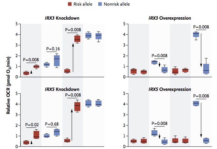
The risk alleles are causative, and mediate through IRX3 and IRX5, because mimicking the repression of IRX3 or IRX5 in 8 carriers of the risk alleles, but not 10 carriers of the non-risk allele increased stimulated metabolism (Fig 3D, shown, left panel). And overexpression of IRX3 or IRX5 reduced stimulated metabolism in non-risk allele carriers (because their endogenous genes are repressed) but not in risk allele carriers (Fig 3D, right panel).

Figure 3D. Oxygen consumption rate (OCR), basal and stimulated, in cells with risk or non-risk alleles.

The take-home messages are that the causative SNP can be kilobases away from the genes that mediate the effect (not a huge surprise) and that small risks might develop from relatively big differences in particular developmental and cell biological pathways (reverse butterfly effect: not small-change-to-big-effect but big effect leads to small change (1-2% BMI)).

N Engl J Med. 2015 Sep 3;373(10):895-907. “FTO Obesity Variant Circuitry and Adipocyte Browning in Humans.” Claussnitzer M, Dankel SN, Kim KH, Quon G, Meuleman W, Haugen C, Glunk V, Sousa IS, Beaudry JL, Puviindran V, Abdennur NA, Liu J, Svensson PA, Hsu YH, Drucker DJ, Mellgren G, Hui CC, Hauner H, Kellis M

Sunday, November 22, 2015

Normalizing T lymphocyte metabolism treats lupus autoimmunity

Glucose is metabolized in two pathways to fuel cellular functions: glycolysis, which splits glucose, yielding little energy but providing pyruvate and other materials for synthesis, and oxidative phosphorylation, which degrades glucose in the mitochondria and produces ~15-fold more energy. Glucose uptake is a limiting in activated T lymphocytes through CD28 costimulation.  Glucose metabolism is dysregulated in T lymphocytes of patients with the autoimmune disease Systemic Lupus Erythematosus (SLE, lupus, review).

These investigators blocked glycolysis with 2-deoxy-D-glucose (2DG) and oxidative phosphorylation with metformin (Met), and observed that disease was reduced and even reversed in mice “triple congenic” (TC) with three lupus-predisposing genetic regions: Sle1-Sle2-Sle3 (review).  2DG is glucose with its 2-hydroxl group replaced by a hydrogen, thereby blocking glycolysis.  Met is a small molecule that was discovered in 1920s to reduce blood glucose, probably by interfering with mitochondrial respiration.  The authors show here that Met reduces extracellular acidification rate (ECAR) and 2DG reduces oxygen consumption rate (OCR), both measures of glucose metabolism, in activated T cells (fig. 1).  


Anti-nuclear antibodies (ANA), a hallmark of lupus, are particularly dangerous because they damage glomeruli, the kidney’s filtration units, causing glomerular nephritis (GN).  The authors show a remarkable reduction in ANA and spleen size (fig. 4, panels, C, D and a portion of panel E shown here) as well as improvement in kidney pathology (fig. 4 panel I)
Although these metabolism inhibitors are not targeted to pathogenic T cells, there are no obvious adverse consequences for the animal or even the immune system.  Treated mice raise antibody responses following protein immunization, generating normal levels and avidities of circulating antibodies (supplemental).  Perhaps the limiting effect of glucose uptake by pathogenic, chronically activated T cells make them more sensitive to inhibition.  How treatment influences control of chronic infections (e.g., EBV, CMV) is also worth knowing.  There was no change in body weight on Met. 
Testing 2 other mouse models of lupus (NZB/W and chronic graft-vs-host (cGVH)), they found a mixture of responses.  For example, in cGVH, combined treatment doesn’t reduce spleens (though Met alone does), while treatment of NZB/W mice reduces ANA but doesn’t improve GN.  Human patients exhibit a range of symptoms and might also be expected to show a range of responses.  This is inspired and inspiring work that cuts across as many disciplines as it does organ systems and raises as many questions as hopes. 
Y. Yin, S.-C. Choi, Z. Xu, D. J. Perry, H. Seay, B. P. Croker, E. S. Sobel, T. M. Brusko, L. Morel, Normalization of CD4+ T cell metabolism reverses lupus. Sci. Transl. Med. 7, 274ra18 (2015).